

Тривимірний біодрук трубчастих структур
Виготовлення кровоносних судин і артерій із справжньої живої тканини за допомогою 3D біопринтера поступово набирає обертів і трансформується в окрему гілку 3D біотехнологій. Тривимірний біодрук активно розвивається, і його застосування у створенні різного роду трубчастих структур стає дедалі популярнішою практикою. Однак його масовому поширенню заважає чимало обмежень, які накладає сам принцип 3D друку.
Звісно, дослідники шукають шляхи обходу наявних труднощів і часом знаходять дуже оригінальні рішення поставлених завдань. Так, команда аргентинських вчених задумала оптимізувати звичайний 3D біопринтер, додавши до нього ще одну, четверту вісь. За словами розробників, тривимірний біодрук на такому пристрої зніме багато обмежень при відтворенні порожнистих, трубчастих структур різної конфігурації.
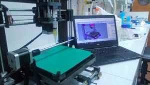
Оптимізований 3D принтер
Унікальна розробка належить команді дослідників з CONICET та Національного університету Ла-Плати під керівництвом доктора Гільєрмо Р. Кастро. Їхньою первісною метою було створення порожнистих трубчастих структур. Стикнувшись зі складнощами та обмеженнями при роботі зі звичайним біодруком, вчені вирішили розробити власну методику відтворення об’ємних виробів. Для цього потрібно було оптимізувати наявне обладнання. У чому суть оригінального покращення?
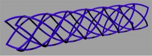
Традиційний тривимірний біодрук відбувається за допомогою стандартних пристроїв із трьома осями XYZ. Ідея дослідників полягає в оснащенні звичайного 3D принтера обертовою четвертою віссю. Насправді це навіть не вісь, а скоріше циліндричне обертове поле побудови. Нанесення матеріалу відбувається безпосередньо на його поверхню. Вісь обертається як за годинниковою стрілкою, так і проти неї, що дозволяє відтворювати різні комбінації структур.
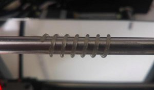
Тривимірний біодрук за новою методикою
Як пояснюють розробники, стандартний FDM 3D друк вимагає нанесення кожного нового шару матеріалу на попередній, що неминуче створює труднощі при отриманні складних елементів. Особливу увагу дослідники приділяють циліндричним, трубчастим і спіральним структурам. Тривимірний біодрук за розробленою методикою дозволяє відтворювати всі необхідні елементи на 3D принтері, обладнаному інжектором зі змінними соплами.
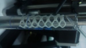
Що стосується матеріалів, команда розглядає застосування різноманітних біополімерних сумішей, що містять альгінат, пектин, хітозан і гідрогель. Залежно від конкретних цілей ці склади змінюються і модифікуються. Зрештою розробники мають намір знайти оптимальний склад для росту живих клітин у 3D-друкованих каркасах. У перспективі така методика може успішно застосовуватися для виготовлення імітації кровоносних судин та інших трубчастих структур людського організму.


 пластик MonoFilament 1.75 мм 0.75 кг Чорний")
 пластик MonoFilament 1.75 мм 0.75 кг Чорний")


Залишити коментар